Tuft Cells
A General Summary
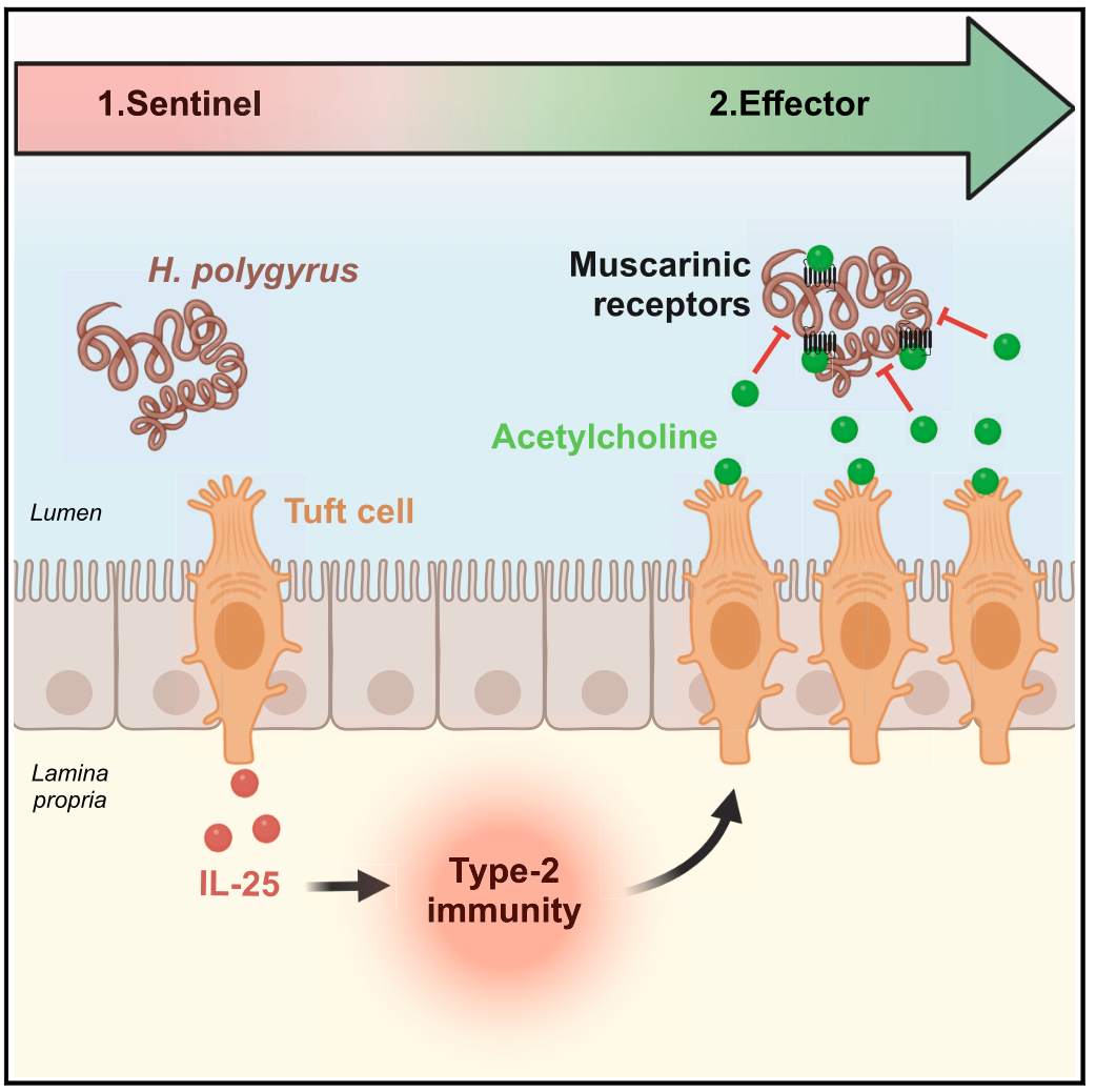
How does the body sense when the gut is invaded by parasites? Tuft cells can “taste” the arrival of worms by sensing their molecular products, and they then alert the immune system by releasing a key messenger molecule termed IL-25. This stimulates nearby immune cells in the body to multiply and induce widespread mobilisation of the immune system. As part of this, tuft cells also multiply and release molecules that directly damage the worm parasites: hence this enigmatic cell type acts both as a sentinel and an executor towards the worms.
In more detail…
Tuft cells are a remarkable subset of cells that fulfil both sensory and effector roles in the immune response to pathogens (Kotas et al 2023). Distantly related to taste bud cells in the tongue, tuft cells are present in epithelial layers of the respiratory and gastrointestinal tracts, expressing G-protein coupled taste receptors (such as Tas2r) that signal through conserved pathways (involving Trpm5).
Intestinal tuft cells are known to play a critical role in immunity to helminth worms, as mice lacking the key transcription factor (Pou2f3) that specifies tuft cell differentiation are unable to expel parasites (Gerbe et al 2016, von Moltke, 2016). Similarly Trpm5-deficient animals fail to generate a tuft cell response to infection (Howitt et al 2016). The nature of the worm substances detected by tuft cells is not yet known, but may include small- and medium-chain fatty acids such as succinic (4-carbon) and undecanoic (11-carbon) acids (Schneider et al 2018; Campillo Poveda et al 2025); it is also possible that they respond to the physical movement of gut worms through mechanosensory receptors.
Once stimulated, tuft cells release the key “alarmin” cytokine IL-25, receptors for which are expressed on innate lymphocyte cells (ILCs) present in the intestinal submucosa. In response, ILCs produce IL-13 for widespread activation of the “type 2” immune response in macrophages, T cells and – most interestingly – intestinal stem cells (Gerbe et al 2016, von Moltke, 2016). Under the influence of IL-13, the stem cells reprogramme to produce more goblet cells (that release worm-trapping mucins) and more tuft cells throughout the intestinal tract.
Gene profiling of tuft cells reveals that they are far more than sentinel cells that raise the alarm; in fact they are instrumental effector cells producing small pharmacologically active molecules that both intensify the immune reaction, and directly attack the parasites. As an example, they express Choline AcetylTransferase (ChAT), the enzyme that converts choline to the neurotransmitter acetylcholine (ACh). Other epithelial cells in the intestinal barrier respond to ACh by increasing ion and fluid export into the lumen of the gut, promoting the expulsion of worms (in what is known as “weep and sweep”) (Billipp et al, 2024). In addition, ACh directly interferes with neurotransmission in the parasitic worms residing in the gut lumen, rendering them unable to resist the “weep and sweep” (Ndjim et al, 2024).
Taken together, tuft cells represent a highly-evolved specialised cell type that guards against worm infection, firstly by expressing receptors that detect invasion, and then by mobilising synthetic machinery to release highly effective anti-worm products in the gut.
[This item was written for the UK Gut Immune Brain Axis Network website]
References
Billipp, T.E., Fung, C., Webeck, L.M., Sargent, D.B., Gologorsky, M.B., Chen, Z., McDaniel, M.M., Kasal, D.N., McGinty, J.W., Barrow, K.A., Rich, L.M., Barilli, A., Sabat, M., Debley, J.S., Wu, C., Myers, R., Howitt, M.R. and von Moltke, J. (2024). Tuft cell-derived acetylcholine promotes epithelial chloride secretion and intestinal helminth clearance. Immunity 57: 1243-1259 e1248. PMC11168877 10.1016/j.immuni.2024.03.023. Link to Publisher’s Website.
Campillo Poveda, M., Loser, S., Gillan, V., Richards, J., Ciancia, C., Blackburn, G., Kerr, E., Barrett, M., Hildersley, K.A., Jay, P., Devaney, E., McNeilly, T.N., Britton, C. and Maizels, R.M. (2025). Metabolomic and functionalanalyses of small molecules secreted by intestinal nematodes in the activation of epithelial tuft cells. Metabolomics 21: 55. PMC12011944 10.1007/s11306-025-02248-w. Link to Publisher’s website.
Feng, X., Fluchter, P., De Tenorio, J.C. and Schneider, C. (2024). Tuft cells in the intestine, immunity and beyond. Nat Rev Gastroenterol Hepatol 21: 852-868. 10.1038/s41575-024-00978-1. Link to Publisher’s website.
Gerbe, F., Sidot, E., Smyth, D.J., Ohmoto, M., Matsumoto, I., Dardalhon, V., Cesses, P., Garnier, L., Pouzolles, M., Brulin, B., Bruschi, M., Harcus, Y., Zimmermann, V.S., Taylor, N., Maizels, R.M. and Jay, P. (2016). Intestinal epithelial tuft cells initiate type 2 mucosal immunity to helminth parasites. Nature 529: 226-230. 10.1038/nature16527. Link to Publisher’s website.
Howitt, M.R., Lavoie, S., Michaud, M., Blum, A.M., Tran, S.V., Weinstock, J.V., Gallini, C.A., Redding, K., Margolskee, R.F., Osborne, L.C., Artis, D. and Garrett, W.S. (2016). Tuft cells, taste-chemosensory cells, orchestrate parasite type 2 immunity in the gut. Science 351: 1329-1333. PMC5528851 10.1126/science.aaf1648. Link to Publisher’s website.
Kotas, M.E., O’Leary, C.E. and Locksley, R.M. (2023). Tuft cells: context- and tissue-specific programming for a conserved cell lineage. Annu Rev Pathol 18: 311-335. 10.1146/annurev-pathol-042320-112212. Link to Publisher’s website.
Ndjim, M., Gasmi, I., Herbert, F., Josephine, C., Bas, J., Lamrani, A., Coutry, N., Henry, S., Zimmermann, V.S., Dardalhon, V., Campillo Poveda, M., Turtoi, E., Thirard, S., Forichon, L., Giordano, A., Ciancia, C., Homayed, Z., Pannequin, J., Britton, C., Devaney, E., McNeilly, T.N., Berrard, S., Turtoi, A., Maizels, R.M., Gerbe, F. and Jay, P. (2024). Tuft cell acetylcholine is released into the gut lumen to promote anti-helminth immunity. Immunity 57:1260-1273 10.1016/j.immuni.2024.04.018. Link to Publisher’s website.
Schneider, C., O’Leary, C.E., von Moltke, J., Liang, H.E., Ang, Q.Y., Turnbaugh, P.J., Radhakrishnan, S., Pellizzon, M., Ma, A. and Locksley, R.M. (2018). A metabolite-triggered tuft cell-ILC2 circuit drives small intestinal remodeling. Cell 174: 271-284 e214. PMC6046262 10.1016/j.cell.2018.05.014. Link to Publisher’s website.
von Moltke, J., Ji, M., Liang, H.E. and Locksley, R.M. (2016). Tuft-cell-derived IL-25 regulates an intestinal ILC2- epithelial response circuit. Nature 529: 221-225. PMC4830391 10.1038/nature16161. Link to Publisher’s website.