Quick Summary : Macrophages and Helminths
Macrophages are intimately involved in the immune response at every stage of helminth infection, adopt specific phenotypes (generally within the Type 2 orbit) and co-ordinate multiple mechanisms to attack parasites and promote expulsion (Coakley et al., 2020).
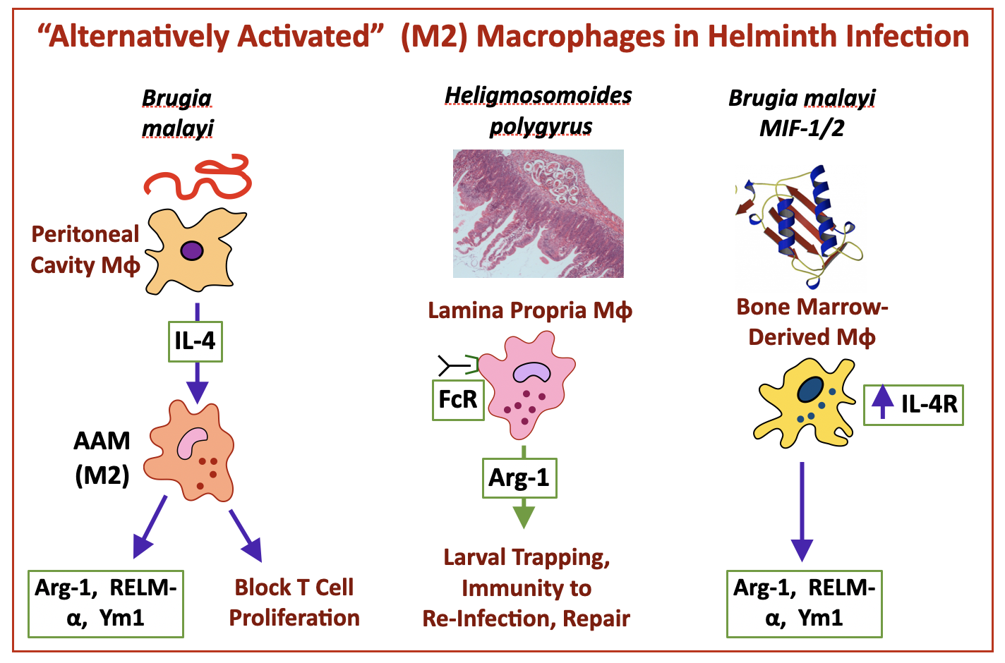
Macrophage properties are strongly conditioned by their tissue environment, with some populations (such as alveolar and enteric muscularis macrophages) predisposed to tolerance induction, and other primed to respond to both host (alarmins, chemokines and cytokines) and parasite (pathogen-associated molecular patterns) signals. In the case of helminth infections the Type 2 (M2) or alternatively-activated macrophage (AAM) phenotype predominates, downstream of alarmin (IL-25, IL-33) signals and IL-4R engagement, to which macrophages respond with production of key mediators such as Arginase-1.
The profile of macrophage responses depends on the tissue location, and on the life history of each helminth infection that may involve skin, lung, serous cavity, submucosal and luminal environments. The Maizels lab has worked on macrophages in Brugia malayi, Litomosoides sigmodontis and Heligmosomoides polygyrus model systems, and how products from these parasites modulate macrophage function.
Macrophages in the Maizels Lab: filarial nematodes Brugia malayi and Litomosoides sigmodontis
Mice are resistant to infection with larvae of the human filarial nematode, B. malayi. However, adult worms survive if transplanted intraperitoneally into mice. Within the infected peritoneal cavity, we discovered the first example of in vivo differentiation of “Alternatively Activated Macrophages” (now more commonly termed “M2”). These potently block proliferation of T cells while sparing their ability to produce cytokines (Allen et al., 1996 and Loke et al., 2000). B. malayi-induced macrophages express key products now recognised as signature genes of the M2 phenotype: Arginase-1, RELM-α (aka Fizz1) and Ym1 (aka Chi3l1) Nair et al 2005.
While resistant to B. malayi, mice can harbour a full infection with L. sigmodontis, that establishes in the pleural cavity and is enabled by regulatory T cells (Taylor et al 2005). Early in infection, prior to release of blood-borne microfilariae (MF), suppressive AAMs are found in the pleural cavity; but following appearance of MF, AAM suppression of T cell proliferation becomes systemic (Taylor et al., 2006). Notably AAM effects can be inhibited with anti-TGFβ antibody, further evidence of the role of that cytokine in helminth infection.
Macrophages in H polygyrus
In infection of mice with H. polygyrus, macrophage responses are skewed to the M2/AAM phenotype, and populate submucosal granulomas around tissue-encysted larvae (as in the cartoon above). Similarly, RELMα+Ym1+ macrophages proliferate and expand in the peritoneal cavity over ther first 7 days of H. polygyrus infection (Jenkins et al., 2012) In susceptible mouse strains such as C57BL/6, larvae grow in the submucosa over 7-8 days and then remigrate as adults back to the intestinal lumen. More resistant strains (BALB/c and SJL) show a stronger granulomatous response with greater effects on larvae which are either trapped in the granuloma, or emerge with reduced fitness and are expelled quickly from the lumen (data from Filbey et al., 2014)
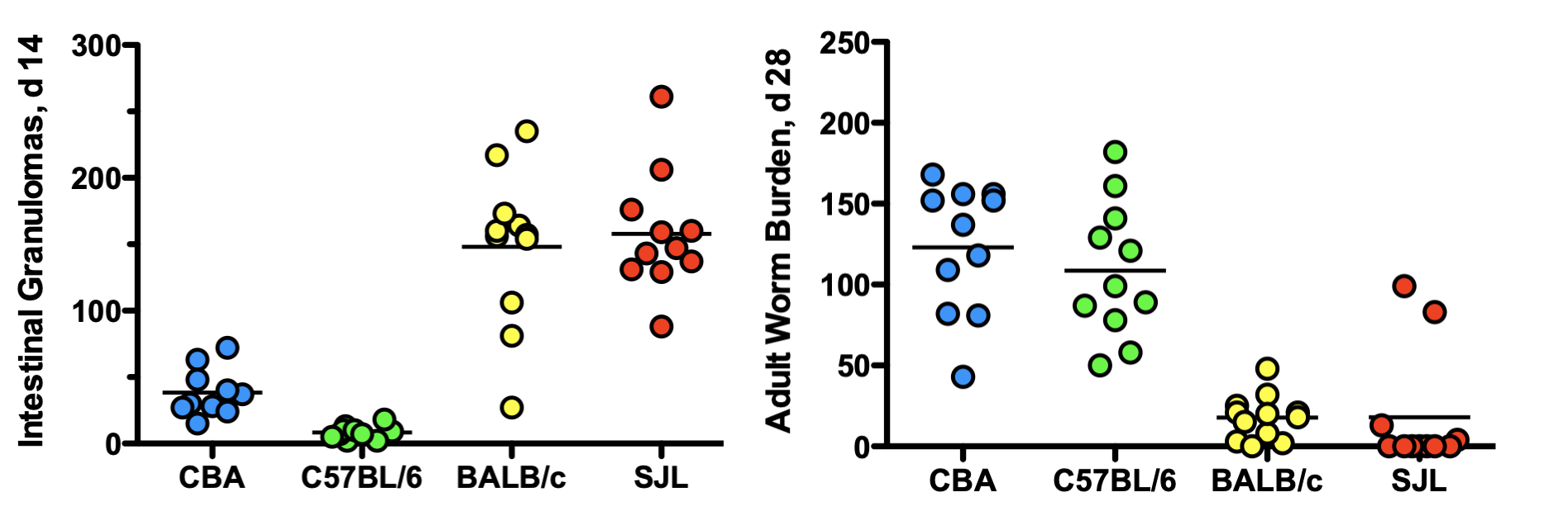
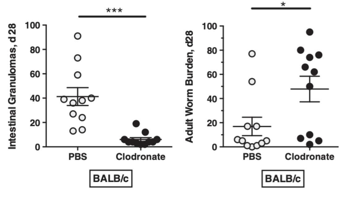
When primary H. polygyrus infections are cleared by drug treatment, mice then display potent immunity to reinfection: this was shown by Anthony et al (2006) ) to depend on the macrophage response as depletion with clodronate abrogated resistance, as did administration of an inhibitor of arginase enzymatic activity. Similarly, clodronate treatment impairs granuloma formation around tissue larvae in BALB/c and SJL mice, with an overall reduction in their ability to expel the worms (Filbey et al., 2014).
Subsequently, the Voehringer lab showed that STAT6 activated macrophages are in isolation insufficient, with epithelial STAT6 signalling also required for expulsion; these authors also showed that in secondary immunity, Arginase-1 was no longer essential indicating multiple redundant mechanisms at play (Westermann 2023).
How do macrophages attack H. polygyrus?
In primary infection Arginase-1 may deplete the environment of this essential amino acid so depriving it from parasites during their growth phase. However, for potent immunity antibody-mediated mechanisms are required, as B-cell deficient mice were unable to clear the worm.: Macrophage activation is promoted by helminth-specific antibodies leading to direct binding to and immobilization of the parasites (Esser-von Bieren et al., 2013; Esser-von Bieren et al., 2015a), with the same phenotype associated with repair of epithelial lesions caused by worm migration (Esser von Bieren et al., 2015b). When mice vaccinated with HES are infected, there is a 4-fold increase in myeloid cell infiltration into infected submucosal tissue, with subsequent trapping of larvae that are unable to return to the lumen (Hewitson et al., 2015).
To expel luminal adult parasites, the host increases fluid and mucus egress into the intestinal tract, while accelerating peristalsis through smooth muscle hypercontractility – which together with constitutes the “weep-and-sweep” response: this process is dependent on signalling to the enteric nervous system by immune cells such as AAMs.
How do helminths counter the macrophage threat?
Helminth parasites release a spectrum of immunomodulators, many of which directly target macrophages (Maizels 2018), including in the case of H. polygyrus, those listed below:
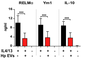
Extracellular vesicles (EVs) released by adult parasites cultured in vitro directly suppress bone marrow-derived macrophage (BMDM) activation, counteracting IL-4/-13-mediated induction of RELM-α, YM-1 and IL-10 (Coakley et al 2017).
Recombinant H. polygyrus TGF-β mimicsTGM1 and TGM4 proteins, corresponding to products in adult HES, block inflammatory LPS-elicted IL-1β, IL-6 and TNF cytokine responses of BMDMs to LPS, and their RELMα response to IL-4 stimulation (Singh et al, 2025).
REFERENCES
Allen, J.E., Lawrence, R.A. and Maizels, R.M. (1996). APC from mice harbouring the filarial nematode, Brugia malayi, prevent cellular proliferation but not cytokine production. International Immunology 8: 143-151. 10.1093/intimm/8.1.143. http://www.ncbi.nlm.nih.gov/pubmed/8671598
Anthony, R.M., Urban, J.F., Jr., Alem, F., Hamed, H.A., Rozo, C.T., Boucher, J.L., Van Rooijen, N. and Gause, W.C. (2006). Memory TH2 cells induce alternatively activated macrophages to mediate protection against nematode parasites. Nat Med 12: 955-960. http://www.ncbi.nlm.nih.gov/entrez/query.fcgi?cmd=Retrieve&db=PubMed&dopt=Citation&list_uids=16892038
Bohnacker, S., Henkel, F.D.R., Hartung, F., Geerlof, A., Riemer, S., Prodjinotho, U.F., Salah, E.B., Mourao, A.S.D., Bohn, S., Teder, T., Thomas, D., Gurke, R., Boeckel, C., Ud-Dean, M., Konig, A.C., Quaranta, A., Alessandrini, F., Lechner, A., Spitzlberger, B., Kabat, A.M., Pearce, E., Haeggstrom, J.Z., Hauck, S.M., Wheelock, C.E., Jakobsson, P.J., Sattler, M., Voehringer, D., Feige, M.J., da Costa, C.P. and Esser-von Bieren, J. (2024). A helminth enzyme subverts macrophage-mediated immunity by epigenetic targeting of prostaglandin synthesis. Sci Immunol 9(102): eadl1467. 10.1126/sciimmunol.adl1467. https://www.ncbi.nlm.nih.gov/pubmed/39642243
Coakley, G. and Harris, N.L. (2020). Interactions between macrophages and helminths. Parasite Immunol 42(7): e12717. 10.1111/pim.12717. http://www.ncbi.nlm.nih.gov/pubmed/32249432
Coakley, G., McCaskill, J.L., Borger, J.G., Simbari, F., Robertson, E., Millar, M., Harcus, Y., McSorley, H.J., Maizels, R.M. and Buck, A.H. (2017). Extracellular vesicles from a helminth parasite suppress macrophage activation and constitute an effective vaccine for protective immunity. Cell Rep 19(8): 1545-1557. PMC5457486 10.1016/j.celrep.2017.05.001. https://www.ncbi.nlm.nih.gov/pubmed/28538175
Esser-von Bieren, J., Mosconi, I., Guiet, R., Piersgilli, A., Volpe, B., Chen, F., Gause, W.C., Seitz, A., Verbeek, J.S. and Harris, N.L. (2013). Antibodies trap tissue migrating helminth larvae and prevent tissue damage by driving IL-4Ralpha-independent alternative differentiation of macrophages. PLoS Pathog 9(11): e1003771. 3828184 10.1371/journal.ppat.1003771
PPATHOGENS-D-13-01662 [pii]. http://www.ncbi.nlm.nih.gov/entrez/query.fcgi?cmd=Retrieve&db=PubMed&dopt=Citation&list_uids=24244174
Esser-von Bieren, J., Volpe, B., Kulagin, M., Sutherland, D.B., Guiet, R., Seitz, A., Marsland, B.J., Verbeek, J.S. and Harris, N.L. (2015). Antibody-mediated trapping of helminth larvae requires CD11b and Fcγ receptor I. J Immunol 194(3): 1154-1163. 4298127 10.4049/jimmunol.1401645. http://www.ncbi.nlm.nih.gov/pubmed/25548226
Esser-von Bieren, J., Volpe, B., Sutherland, D.B., Burgi, J., Verbeek, J.S., Marsland, B.J., Urban, J.F., Jr. and Harris, N.L. (2015). Immune antibodies and helminth products drive CXCR2-dependent macrophage-myofibroblast crosstalk to promote intestinal repair. PLoS Pathog 11(3): e1004778. 4373753 10.1371/journal.ppat.1004778. http://www.ncbi.nlm.nih.gov/pubmed/25806513
Filbey, K.J., Grainger, J.R., Smith, K.A., Boon, L., van Rooijen, N., Harcus, Y., Jenkins, S., Hewitson, J.P. and Maizels, R.M. (2014). Innate and adaptive type 2 immune cell responses in genetically controlled resistance to intestinal helminth infection. Immunol Cell Biol 92: 436-448. PMC4038150 10.1038/icb.2013.109. http://www.ncbi.nlm.nih.gov/pubmed/24492801
Hewitson, J.P., Filbey, K.J., Esser-von Bieren, J., Camberis, M., Schwartz, C., Murray, J., Reynolds, L.A., Blair, N., Robertson, E., Harcus, Y., Boon, L., Huang, S.C., Yang, L., Tu, Y., Miller, M.J., Voehringer, D., Le Gros, G., Harris, N. and Maizels, R.M. (2015). Concerted activity of IgG1 antibodies and IL-4/IL-25-dependent effector cells trap helminth larvae in the tissues following vaccination with defined secreted antigens, providing sterile immunity to challenge infection. PLoS Pathog 11(3): e1004676. 4376884 10.1371/journal.ppat.1004676. http://www.ncbi.nlm.nih.gov/pubmed/25816012
Jenkins, S.J., Ruckerl, D., Thomas, G.D., Hewitson, J.P., Duncan, S., Brombacher, F., Maizels, R.M., Hume, D.A. and Allen, J.E. (2013). IL-4 directly signals tissue-resident macrophages to proliferate beyond homeostatic levels controlled by CSF-1. J Exp Med 210(11): 2477-2491. jem.20121999 [pii]
10.1084/jem.20121999. http://www.ncbi.nlm.nih.gov/entrez/query.fcgi?cmd=Retrieve&db=PubMed&dopt=Citation&list_uids=24101381
Loke, P., MacDonald, A.S., Robb, A., Maizels, R.M. and Allen, J.E. (2000). Alternatively activated macrophages induced by nematode infection inhibit proliferation via cell to cell contact. European Journal of Immunology 30: 2669-2678.
Maizels, R.M., Smits, H.H. and McSorley, H.J. (2018). Modulation of host immunity by helminths: the expanding repertoire of parasite effector molecules. Immunity 49(5): 801-818. PMC6269126 10.1016/j.immuni.2018.10.016. https://www.ncbi.nlm.nih.gov/pubmed/30462997
Nair, M.G., Gallagher, I.J., Taylor, M.D., Loke, P., Coulson, P.S., Wilson, R.A., Maizels, R.M. and Allen, J.E. (2005). Chitinase and Fizz family members are a generalized feature of nematode infection with selective upregulation of Ym1 and Fizz1 by antigen-presenting cells. Infection and Immunity 73: 385-394. http://www.ncbi.nlm.nih.gov/entrez/query.fcgi?cmd=Retrieve&db=PubMed&dopt=Citation&list_uids=15618176
Taylor, M., Le Goff, L., Harris, A., Malone, E., Allen, J.E. and Maizels, R.M. (2005). Removal of regulatory T cell activity reverses hyporesponsiveness and leads to filarial parasite clearance in vivo. Journal of Immunology 174: 4924-4933.
Taylor, M.D., Harris, A., Nair, M.G., Maizels, R.M. and Allen, J.E. (2006). F4/80+ alternatively activated macrophages control CD4+ T cell hyporesponsiveness at sites peripheral to filarial infection. J Immunol 176: 6918-6927. http://www.ncbi.nlm.nih.gov/entrez/query.fcgi?cmd=Retrieve&db=PubMed&dopt=Citation&list_uids=16709852
Westermann, S., Schubart, C., Dietschmann, A., Castiglione, K., Radtke, D. and Voehringer, D. (2023). Th2-dependent STAT6-regulated genes in intestinal epithelial cells mediate larval trapping during secondary Heligmosomoides polygyrus bakeri infection. PLoS Pathog 19(4): e1011296. PMC10109486 10.1371/journal.ppat.1011296. https://www.ncbi.nlm.nih.gov/pubmed/37018382